
Every time we collect the plastic from the ocean deployments, we marvel at the thickness of the biofilm attached to it, laugh at the little crabs running around, and just ooh and awe at life using any surface to grow and expand. We can see it, and we can feel it- especially when the cages need to be scrubbed and washed in a sonicated bath before reuse. But there is a special appreciation when we can actually “see it” at the microscopic level thanks to technology, in this case scanning electron microscopy (SEM).
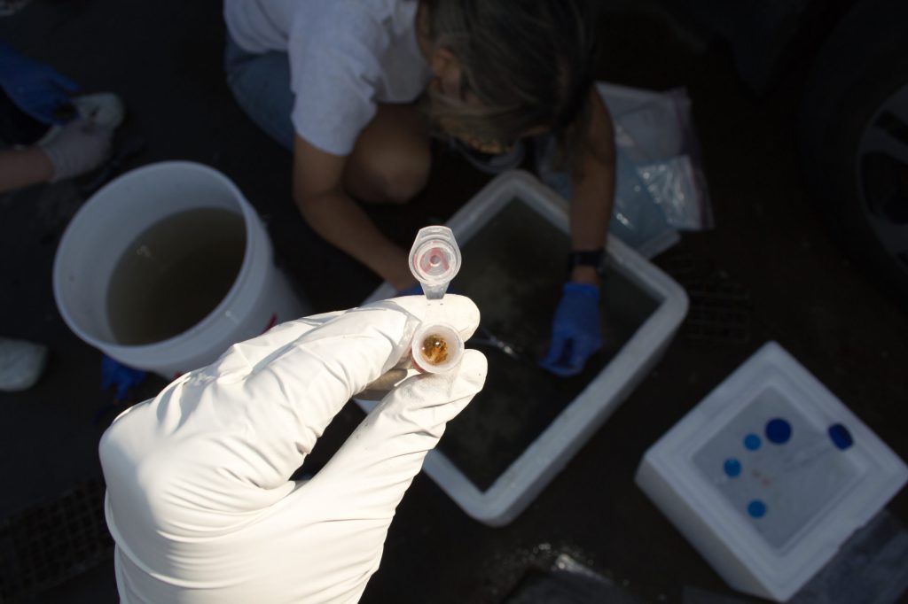
Last time we collected samples back in December we planned for SEM in coordination with The Scripps Research Institute (TSRI) Microscopy Core. It was a chore- EM in general is finicky and samples have to be handled in a way that it preserves morphology. That meant not exposing the samples coming from underwater to air at any time. The logistics of the process were a bit daunting, and were solved by using a big “Ace hardware” bucket full of ocean water to haul the cages from the divers to our processing station. The TSRI people made the fixative using ocean water, and Rachel and I were elbow deep in the water opening cages and cutting the plastic to get them in the fixative. Our prospective research student Andrea Price was there, thankfully! She helped both with organizing the tubes and taking great pictures of the process.

Samples were then dropped off at TSRI and the wait began. The sampling happened just before Christmas so it took a while. But yesterday we finally had our SEM watching session (via zoom, of course). Dr. Kim Vanderpool walked us through the samples and the controls, and we spent a few hours looking for organisms to take pictures of. The one below is from plastic #4 (a typical grocery bag plastic) after 53 days. We called the large things “diatoms,” Emelia said they could be coccolithophores. I claim ignorance in knowledge of algae 🙂 On the other hand it was fun to see all the bacteria in-between and on top, of all kinds of shapes and forms.

This is just one sample- we have pictures of three plastics, both control and samples, at different magnifications. What it shows is something we knew macroscopically- that there is a huge biological richness to the plastisphere even after a relatively short incubation. Most SEM images have come from microplastics that have been in the water for much longer time, allowing the plastic to break down to tiny pieces. We are looking forward the next DNA results, in which we’ll be able to compare colonization by means of a surface only (glass slides) versus by attraction to plastic.
But yesterday was a fun day. One of those that left us with a warm fuzzy inner glow. Scientists need it once in a while!
